Hôpital Privé de Bois Bernard
Prise de RDV: 03 21 79 26 56
Secrétariat: 03 21 79 26 36

Cette page est dédiée aux Professionnels de Santé
Quels Examens prescrire?
Les radiographies standards: Obligatoires
Une échographie?
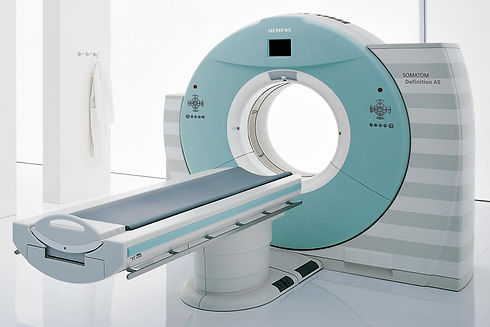
Un Arthroscanner et/ou une IRM?

Les radiographies standards
Face Profil bilan de base
Profil de Lamy bilan de base
Humérus 3 rotations recherche de calcifications
-
Face Rotation Neutre
-
Face Rotation Externe
-
Face Rotation Interne
Profil de Bernageau Instabilité épaule
Incidence de Garth Instabilité épaule

Face Rotation Externe. Face Rotation Neutre Face Rotation Interne

1. Bord antéro inférieur de la glène.
2. Bord antéro supérieur de la glène.
3. Bord postérieur de la glène.
4. Acromion.
5. Processus coracoïde.

1.Scapula vue de profil.
2. Espace sous acromial.
3. Diaphyse humérale.
4. Clavicule.
5. Acromion.
6. Processus coracoide
L'échographie
-
Examen très dépendant du Radiologue
-
Examen de débrouillage
-
Ne suffit pas de façon isolée
-
Examen intéressant dans le but d’une exploration dynamique
-
Instabilité du biceps
-


L'arthro-scanner
-
Examen de choix
-
Précise la rupture de coiffe (taille et siège)
-
Précise une rupture en face profonde (Parfois traumatique)
-
Précise l’état des muscle (dégénérescence graisseuse) (Classification de Goutallier)
-
Précise l’état et la position du chef long du biceps
-
Excellente exploration du bourrelet
-
Recherche de lésions labrales
-
Recherche de SLAP lésions
-
-
Évaluation du stock osseux glénoïdien (Faisabilité d’une arthroplastie d’épaule)
-
DEFAUTS :
-
ne précise pas l’existence d’une tendinite isolée
-
Ne précise pas une bursite de l’espace sous acromial
-
N’explore pas la face Bursale de la coiffe (Face superficielle) (Faux négatif pour une rupture partielle superficielle de la coiffe = Rupture non transfixiante)
-
Le Burso-scanner explore les lésions superficielles (Examen rare)
-
-
-
LIMITES
-
Intolérance à l’iode
-
Patients diabétique ou insuffisant rénal
-
L'IRM
-
Intérêt ++ si conflit sous-acromial isolé (bursite sous acromiale)
-
Examen de choix si coiffe inflammatoire (Tendinite ou Bursite)
-
Précise la rupture de coiffe (taille et siège)
-
Précise l’état des muscles (dégénérescence graisseuse) (Moins performante que l’arthroscanner)
-
Précise l’état et la position du chef long du biceps
-
Exploration du bourrelet (Moins performante que l’arthroscanner)
-
Très bon examen pour quantifier le caractère inflammatoire d’une arthropathie acromio-claviculaire
-
Evalution du stock osseux glénoïdien (Faisabilité d’un arthroplastie d’épaule)
-
Kystes spino-glénoidiens
-
Capsulites (Mais le diagnostic est clinique)
-
Précise l’épanchement bursal
-
Précise une rupture partielle (en Face superficielle)
-
DEFAUTS :
-
Faux positifs de Ruptures de Coiffe
-
Parfois confusion entre une rupture et une tendinite importante
-
-
LIMITES
-
Claustrophobie du patient
-
Patients porteurs d’implants métalliques (Pace-maker, Clips vasculaires, etc…)
-

Choix de l’Examen / au diagnostic suspecté

Rupture de la coiffe: Arthro-scanner et/ou IRM ?

Il faut parfois y penser...
Lorsque le diagnostic clinique n'est pas cohérent avec l'imagerie
-
Après avoir éliminer une Capsulite Rétractile (Examen clinique +++)
-
Devant une épaule déficitaire ou pseudo-paralytique, et/ou avec une amyotrophie des fosses supra et infra-épineuses, ou du deltoïde…
-
Éliminer une cause neurologique
-
Névralgie cervico-brachiale, ou une compression radiculaire, ou médullaire
-
Penser au syndrome de Parsonnage Turner
-
Demander un avis neurologique…et
Un ELECTROMYOGRAMME du membre supérieur
-
Accessoirement un bilan du rachis cervical
-
Radiographies du rachis cervical
-
IRM médullaire cervicale
-
-
Devant une épaule Hyper-algique …
Une scintigraphie
-
Éliminer une algo-neurodystrophie
-
Doute sur l'organicité des symptômes (sinistrose?)
-
Doute sur une étiologie infectieuse
-
Doute sur une localisation secondaire...
-
Très rarement
-
Une Arthro-IRM
-
Un Burso-scanner
-
Si difficulté à poser le diagnostic d’une rupture de la face superficielle
-
Et que l’IRM est contre-indiquée…
-